Site Content
HIstory of Microscopy
Viewing live blood under a microscope is probably as old as the microscope itself. But it was the work of European scientists Dr. Antoine Bechamp and Dr. Gunther Enderlein in the mid-19th and early 20th Centuries that would advance the use of the microscope, challenge the medical establishment of the day and propose new ways of interpreting what was being viewed in blood. Other microscopists included noted physiologist, Dr. Claude Bernard, who coined the term "internal milieu," Germ Theory advocate Louis Pasteur, Californian Dr. Virginia Livingston Wheeler and Canadian scientist Gaston Naessens. In the 1920s European medical practitioners added another twist to unconventional microscopy when they began looking at dried blood samples, later called the Oxidative Stress Test. A glass microscope slide is dabbed onto a bead of blood on the finger in sequence several times, resulting in a slide with 8 individual drops of blood pressed upon the slide and allowed to air dry. The resulting patterns seen in the dry blood under the bright field format reveal a characteristic "footprint" which can be seen in similar cases and, thus, are predictive of certain generalized pathologies. For instance, cases of advanced degenerative disease show very poor clotting, minimal fibrin formation with many white "puddles" disseminated throughout the sample. A healthy control subject's blood shows a tight, fibrin rich clotting pattern with no white puddles. In the 1930s, the head of surgery at Massachusetts General Hospital, Dr. H.L. Bowlen, MD, introduced the dry blood test to America. Dr. Bowlen learned the dry test from President Dwight D. Eisenhower's physicians, Drs. Heitlan and LaGarde. In the 1970s, one of Heitlan-LaGarde's students, Dr. Robert Bradford of the American Biologics Hospital in Mexico, began teaching other practitioners how to perform this test. So now there is over 70 years of dry blood testing data by hundreds of health care practitioners worldwide. Nutritional Microscopy is now an alternative examination routinely utilized by holistic medical, osteopathic, chiropractic and naturopathic physicians, as well as other health care professionals around the world, providing an insightful view of the biological terrain.
What is Nutritional Microscopy
Microscopy is not meant to treat or diagnose. It is an educational tool which offers a different look into what is going on within the body. When done correctly, blood analysis doesn’t lie.
There are practitioners who either do not know what they are doing or are outright frauds. Unfortunately, this gives microscopy a bad name. There are many videos online or websites where the practitioners will show the edge of the slide where the blood is dried and coagulated. Then they take a second sample and claim improvements after taking supplement X or procedure Y. In realty all they really did was move to the middle of the slide where they should have been looking all along. There is a video online where the practitioner says, “I have never seen blood move this fast,” while he is actually moving it diagonally with the stage of the microscope. I remember listening to one practitioner claim he had been doing microscopy for x number of years and had seen x thousands of people. When you do the math, he would have had to have seen 21 people a day, 365 days a year for years. Highly unlikely, Be careful out there!
I can think of two striking instances where the truth in the blood stood out.
A 17-year-old girl is brought to see me by her mother. She is 5’ 7” and 80 pounds at best. Her mother says she has an eating disorder and is bulimic. She has even been institutionalized. The girl tells me there is nothing wrong with her. I look at the blood and the girl had a massive parasite profile. After further testing they found out she had a tape worm, not an eating disorder!
A Naturopath and Acupuncturist that I had worked with extensively brought a woman for me to see. The doctors said I would never figure out what was wrong with her and sat in on the session. This poor woman had been to countless doctors, and spent a enormous sums of money over the years looking for answers. When I finished, I simply said to the client, your blood does not match what you’re telling me your symptoms are. The doctors started laughing and said we told you so. . I explained to the woman that the blood does not lie. I asked her what was going on in her head? The doctors looked at me like I had something going on in my head. I sat there in silence and looked into her eyes, within 5 minutes she was crying her eyes out. Her hidden emotions were making her physical ill. The three of us played therapist for the next 45 minutes. Microscopy is an amazing tool when used properly.
• Utilizes a high-powered microscope to view a drop of capillary blood from a subject’s fingertip, obtained with a sterile lancet.
• Examines blood cells and plasma to gather research data: How does the blood picture relate to the level of health challenges experienced by the subject; What are the subjects nutritional needs and/or deficiencies?
• Addresses areas of imbalance suggested by the case history and the blood picture, N. Microscopy is an evaluation of the internal environment referred to as the biological terrain but is not considered diagnostic.
• No medical test by itself is usually considered diagnostic without corroborating lab tests, imaging studies, or physical examination. dfsf Where standard laboratory blood tests are generally quantitative (how many cells are there?), N. Microscopy is qualitative (what is the condition of the cells?). Standard laboratory tests are often used as pre/post studies to N. Microscopy because there is correlative value in knowing both the quantity and quality of the client's cells. The Microscopist and client can see the characteristics of the client’s blood live on a video screen. This process gives current and past information, as it pertains to the biological terrain of the client (stress appears in the blood sometimes years before it manifests as symptoms). This information can assist the Microscopist and client by:
• Giving early warning of possible upcoming challenges
• Showing patterns of disorganization
• Alerting to the advisability of medical referral
• Monitoring a challenge before and after regimes
• Determining the effectiveness of various regimes
OBSERVATION AND MONITORING OF METABOLIC FUNCTION or DYSFUNCTION:
The Phase Contrast (Unchanged Live Blood) and Bright Field (Dry Blood/ Oxidative Stress) Demonstrations are used to observe and monitor metabolic function or dysfunction, thereby taking the guesswork out of diet determination and the selection of appropriate supplementation. Your Wellness Consultant has been trained by the Robert O. Young Research Center to observe the blood for imbalances in body chemistry. In the New Biology these “imbalances” are seen as “conditions” brought on by acidity from poor diet, nutrition, or lifestyle choices.
Among the phenomenon observed are:
• Relative level of acidity in the body fluids and the effects these acids have on the body
• Relative activity of the immune system
• Condition of the red blood cells and changes in form and function
• General organ “stress”
• Presence of parasites, bacteria, yeast, fungus, and mold
• Blood sugar imbalances
• Malabsorption of fats, proteins, and other nutrients
• Crystalline forms of morbid matter, acids, cholesterol, and mycotoxins
• Degenerative stress and gastrointestinal tract dysfunction
Live Blood and Dried Blood analysis

Live Blood Analysis looks at the quality of the blood. This is different than a traditional blood test, which you get from your doctor. They are interested in quantitative information. How much and how many cells, etc. We are more interested in the qualitative information or the condition of what is there. Although the count is important, it doesn’t matter if you have the correct number of cells if they are fermenting and disorganizing.This is what is perceived to be normal health: the shapes of the cells are all even in size and color; the cells are separated from each other and residing in their own space.

Because the live blood has so many variables based on what you ate or drank recently, we also correlate what we see to a second kind of research known as the Mycotoxic
Oxidative Stress Test. These are just big words for where acid is settling in the body. This is what is perceived to be normal health. It is tightly organized and bright red with these black cobweb like rivers running through. The images of the healthy live and dried blood are my own.

Dried Blood Analysis shows a lifetime of challenges. What we do with the dried blood is let it sit on the finger for 30 seconds then pick it up in a series of drops. The bigger drops show things that are on the surface, or recent and as we go farther down we can see things over a life time. Just because we see something doesn't mean you will experience it. It just shows a weakened area in the body.

Where the pools or lakes are in the drop indicates where it is in the body. If the pool is in the center of the drop then the challenge is in the middle of the body. If it is in the edge then its an extremity. If it goes all the way around it is a system. Some of what can be observed are;

During a live blood session we will be able to see if there are bacteria, yeast, mold, enough good fats and oils, premature clotting, acid crystallizations, or any disorganization at the cellular level. This is a picture of a 350- pound woman's blood. She had headaches, diabetes, high blood pressure, arthritis, fibromyalgia, and much more. By far the worst blood I have ever seen.

Fibrin “Nets” and Fibrin “Trees” An advanced stage of fibrin organization associated with a focal point of yeast or bacteria as an intelligent protective mechanism against janitorial effects of White Blood Cells (garbage collectors). Also indicates a high level of latent tissue acidosis. Dr Alan Macdonald refers to this as the cyst form in Lyme Disease. The spirochetes hide in this protective colony. They are associated with joint pain, and arthritis. In someone's blood with medically diagnosed Lyme these will be the predominant profile in the blood.

Rouleax ” Stacking or chaining of red blood cells (RBCs) is related to poor protein metabolism and altered pH or acid imbalance, which varies the electrical negative charge of the cell membrane causing them to stick together. Rendering red blood cells unable to pass into the small capillaries to transfer oxygen or remove CO2. Rouleax is typically caused by acidic beverages, like coffee or soda. It can also be dehydration. It is unusual to see someone's blood that is constantly in a state of Rouleax.

Macrocytes – Microcytes - Ovalcytes (“Anisocytosis”) RBCs are smaller or larger than normal. indicates the ingestion of an excessive amount of over acidic food and drinks which causes a deficiency of sodium bicarbonate in the alkaliphile glands and a compromise of the alkaline pH of the small intestine (7.8 to 8.4). Modern medicine does not pay a lot of attention to the quality of the blood. A few years ago, there was a study that showed if there is a big difference in the size of your cells you can have a increased risk of heart disease. It shows up on the Red Blood Cell Width on traditional blood tests.

Heterogeneous Symplasts This is an organization or grouping of several stages of microzymian organization including yeast, bacteria and fibrin which are highly disruptive to normal blood circulation; indicated in advanced stages of latent tissue acidosis. You will see these in someone with Lyme who is experiencing neurological issues. Also seen in Bells Palsy, MS, and Lupus. I have seen these the size of 14 screens in someone with severe MS. Every child I have ever seen with Autism has had these in their blood. Clean it up and watch the child improve.

Yeast (Y-form, M-form, G-form) Born out of red blood cells ( RBCs) due to blood pH imbalance from latent tissue acidosis; diet too high in protein, carbohydrates/sugars; may be caused by excess antibiotic use, hormonal therapy, steroid use; fungal infections. When you see large quantities of yeast as soon as you start looking at the blood there is cause for concern. Yeast has many different looks. Based on the looks it can point you to the illness or soon to be illness. I sat with a pathologist from a major hospital in Boston. When I showed him images of yeast and told them what they were experiencing he was dumfounded. His response was "I have been doing this for 20 years. I work with 9 other pathologists. None of us would see this." Study finds 80% increased risk of breast cancer from dairy milk.

Erythrocyte (RBC) Aggregation Is due to loss of the negative surface charge; where plasma acids act as molecular glue, causing RBCs to stick together. This results in poor circulation that leads to cold hands, cold feet, increased body temperature, sweating, hot flashes, water retention, bloating, lightheadedness, dizziness, and muddled thinking. When our red blood cells circulate through our capillaries, they have t go single file. This profile creates traffic jam resulting in the above symptoms.

Heavy Metals Appear as dark ring around the outside of sample or black ‘chunks’ or waves spinning to the outside. Perceived to be holding metals in the tissue which may be due to first or secondhand cigarette smoke, environmental pollutants, cleaning products, water pipes. I have worked with a lot of practitioners who test for heavy metals. Clients come and see with their reports showing extremely high levels of mercury in their body. Unfortunately, I have never been able to corelate it with what I see in the blood. The above image is a former pro athlete’s blood. He has been on the cover of Men’s Health and about as fit as you can be. He runs on the beach in LA every day. The dark ring on the edge is from environmental exposure.

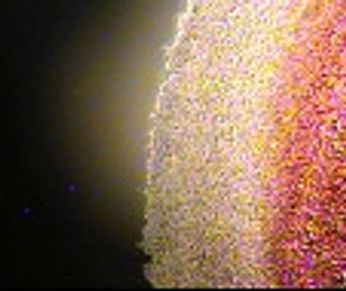
Back, Neck, Shoulder Challenges or Scar Tissue Appear as curved elongated fibrin strands. Perceived to be whiplash, headaches, scoliosis, injury, trauma, subluxation; surgery or injury related scar tissue. Photo is a scar from C-Section.

Liver Stress Appear as black circles around protein pools slightly off center. Perceived to be from toxicity not being removed effectively. Causes are Tylenol, Advil, Aleve, Medication, Hepatitis, and even novocaine. Image is someone with medically diagnosed Hepatitis.

Bowel Toxicity Appears as a dark center in the sample and/or a cluster pattern of protein pools between 10-40 microns. Perceived to be small and large bowel holding toxins, possible damage to the intestinal villas, irregular elimination, and poor digestion with gas, pain or bloating. Picture is of someone ith medically diagnosed Crohn's Disease.

Endocrine Imbalance Appear as a raised area with erased fibrin at outer edges. Perceived to be that the thyroid, parathyroid, and/or pancreas are out of balance. I find it interesting that if someone is on a Thyroid medication and the dosage is correct you do not see a endocrine profile. If the dosage is too high or too low the profile shows up.

Low Alkaline Buffers Mineral Deficiencies Appear as a double coastline or beach. Perceived to be low alkalophile buffers including sodium bicarbonate and minerals. If you use the edge of the red blood as a guide and move it over to the right side of the screen, most of the population is about 1/3rd of a screen. If the beach reaches 3/4 of a screen someone will have heartburn, acid reflux, or indigestion occasionally. If it reaches a full screen, they will have it consistently. Once it is over a full screen someone may be lactose intolerant, or have irritable bowel issues. All are easily fixable.

Hypercalcemia - Mineral Deficiency Appears as white radial spokes (lines) from the center of sample. Perceived to be too much calcium in blood being leached from bones and vital organs to neutralize acid; latent tissue acidosis, lack of alkaline buffers, possible electrolyte imbalance, altered blood pH, possible thyroid/ parathyroid out of balance This profile is like the Ace of Spades. It will trump every other profile and be the only profile visible until it is resolve. This picture is from someone with severe osteoporosis.
Copyright © 2024 Sean Fuller - All Rights Reserved.
Powered by GoDaddy
This website uses cookies.
We use cookies to analyze website traffic and optimize your website experience. By accepting our use of cookies, your data will be aggregated with all other user data.